Introduction
Magnetic Resonance Imaging (MRI) is an invaluable tool in the diagnosis and management of various medical conditions, including sarcomas. Sarcomas are rare, aggressive tumors that originate in the soft tissues or bones. Accurate imaging is crucial for the early detection, characterization, and treatment planning of these tumors. Two important MRI techniques, T1-weighted (T1) and T2-weighted (T2) imaging, play distinct roles in sarcoma evaluation. In this article, we explore the differences between T1 Vs T2 MRI in sarcoma imaging and discuss when each technique is most appropriate.
Understanding T1 and T2 MRI
Before delving into their applications in sarcoma imaging, let’s briefly review T1 and T2 MRI:
T1 MRI:
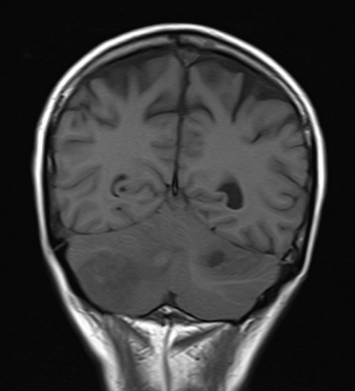
T1-weighted images provide excellent anatomical detail and tissue differentiation.
Tissues with short T1 relaxation times appear bright, while those with long T1 relaxation times appear dark.
Fat has a short T1 and appears bright on T1-weighted images.
T1 MRI is typically used for anatomical assessment and identifying fat-containing structures.
T2 MRI:
T2-weighted images highlight differences in water content and soft tissue contrast.
Tissues with long T2 relaxation times appear bright, while those with short T2 relaxation times appear dark.
Fluids and edematous tissues have long T2 times and appear bright on T2-weighted images.
T2 MRI is often used to evaluate tissue characteristics, inflammation, and tumor extent.
T1 MRI in Sarcoma Imaging
T1-weighted MRI is essential for sarcoma imaging in several scenarios:
Tissue Characterization: T1-weighted images help differentiate between fat and other soft tissues. Since some sarcomas contain areas of fat, T1 MRI can assist in distinguishing benign lipomas from potentially malignant liposarcomas.
Pre- and Post-Contrast Imaging: T1-weighted images are commonly acquired before and after the administration of a gadolinium-based contrast agent. This allows for the assessment of tumor vascularity and enhancement patterns, aiding in tumor grading and treatment planning.
Anatomical Localization: T1 MRI provides high-resolution anatomical images that are crucial for identifying the exact location and extent of sarcomas. This information is essential for surgical planning and radiation therapy.
Monitoring Treatment Response: T1-weighted images can be used to assess the response of sarcomas to treatment, such as chemotherapy or radiation therapy, by measuring changes in tumor size and contrast enhancement.
T2 MRI in Sarcoma Imaging
T2-weighted MRI is equally important for sarcoma evaluation:
Tumor Characterization: T2-weighted images provide information about the water content within tissues, helping in characterizing the composition of sarcomas. High T2 signal within a tumor may suggest necrosis or cystic components
Differentiating Tumor from Surrounding Tissues: Sarcomas often infiltrate adjacent tissues. T2 MRI can help delineate the tumor’s extent and distinguish it from normal tissues, aiding in surgical planning.
Assessing Edema: T2-weighted images are valuable for detecting peritumoral edema, which can impact treatment decisions and prognosis. Increased T2 signal in the surrounding tissue may indicate a more aggressive tumor.
Evaluating Response to Therapy: T2 MRI is useful for monitoring changes in tumor characteristics following treatment. Reduction in peritumoral edema or cystic areas may indicate a positive response to therapy.
Choosing the Right MRI Technique
The choice between T1 and T2 MRI in sarcoma imaging depends on the clinical context and the specific information needed:
For initial assessment, localization, and anatomical detail, T1 MRI is often the first choice.
When characterizing tumor composition, assessing edema, or monitoring changes in tumor characteristics over time, T2 MRI is indispensable.
Combining both T1 and T2 MRI sequences, along with other specialized sequences like diffusion-weighted imaging (DWI) and post-contrast imaging, provides a comprehensive evaluation of sarcomas.
Conclusion
T1 VS T2 MRI are complementary tools in the imaging of sarcomas. While T1-weighted images excel in providing anatomical information and assessing tumor vascularity, T2-weighted images offer insights into tumor composition, edema, and changes following treatment. The judicious use of these techniques, often in combination, enables accurate diagnosis, staging, treatment planning, and monitoring of sarcomas, ultimately improving patient outcomes. The choice between T1 and T2 MRI depends on the specific clinical questions and the information needed for optimal patient care.

